
Ritrattamento reendodontico con perforazione radicolare apicale del dente 11: relazione di caso scientifico accademico.
Traduzione automatica
L'articolo originale è scritto in lingua EN (link per leggerlo) .
Abstract
La perforazione radicolare si riferisce a una comunicazione patologica tra il sistema canalare radicolare e la superficie esterna del dente. Questa complicazione può sorgere a causa di vari fattori, tra cui carie estese, riassorbimento radicolare o uso inappropriato di strumenti endodontici. La scelta dei materiali utilizzati durante il trattamento è fondamentale per sigillare efficacemente l'area interessata. Questi materiali devono possedere specifiche caratteristiche, come biocompatibilità, capacità di sigillatura, solubilità in acqua, non citotossicità e la capacità di promuovere la riparazione e la rigenerazione dei tessuti. Di conseguenza, i cementi bioceramici riparativi sono stati riconosciuti come agenti idonei per affrontare questa forma di lesione. L'obiettivo principale di questo studio è presentare un rapporto di caso clinico che dimostri l'uso efficace del cemento bioceramico riparativo nel sigillare una perforazione radicolare durante il ritratamento del dente 11.
Introduction
Gli incidenti chirurgici possono rappresentare sfide significative durante la terapia endodontica e possono contribuire al fallimento del trattamento. Vari fattori possono predisporre a questi incidenti durante la pulizia del canale radicolare, tra cui calcificazione, noduli pulpari, denti malposizionati nell'arcata dentale, carie estese, riassorbimento radicolare e perforazioni radicolari.
La perforazione della radice è caratterizzata da una comunicazione tra il canale radicolare e la superficie esterna del dente. Questo tipo di lesione iatrogena può portare a diverse conseguenze, che vanno dall'infiammazione cronica nel parodonto con tessuto di granulazione alla perdita ossea alveolare.
Esiste una gamma di materiali disponibili per il trattamento delle perforazioni radicolari, come amalgama, idrossido di calcio, ionomero di vetro, aggregato di triossido minerale (MTA) e materiali di riparazione bioceramici. L'MTA, contenente silicato di tricalcio, alluminato di tricalcio, ossido di tricalcio e ossido di silicato, è utilizzato non solo per le perforazioni radicolari ma anche per la copertura diretta della polpa e il riempimento retrogrado. Offre diversi vantaggi, tra cui biocompatibilità, azione batteriostatica e buona capacità di sigillatura.
I materiali bioceramici, come descritto da Raghavendra et al., sono cementi di riparazione biocompatibili noti per il loro elevato potenziale di sigillatura e le proprietà antimicrobiche. Questi materiali, comunemente utilizzati in odontoiatria, comprendono silicato di calcio, fosfato di calcio, zirconia e vetri bioattivi, e sono classificati come cementi a base di silicato di calcio o cementi idraulici a base di silicato di calcio.
Sousa et al. menzionano che questi materiali di riparazione sono sintetizzati attraverso vari processi chimici, il che conferisce loro un'eccellente biocompatibilità e la capacità di indurre una risposta rigenerativa nei tessuti assorbendo sostanze osteoinduttive durante il processo di guarigione ossea. Sono stati introdotti in odontoiatria principalmente per la riparazione e l'otturazione dei canali radicolari.
Sebbene nessun materiale possieda tutte le proprietà ideali per trattare le perforazioni radicolari, i cementi di riparazione bioceramici hanno guadagnato una notevole popolarità grazie ai loro eccellenti risultati di sigillatura a lungo termine. Pertanto, questo studio mira a dimostrare la sigillatura riuscita di una perforazione radicolare con cemento di riparazione bioceramico durante il ritratamento della terapia endodontica nel dente 11 attraverso la presentazione di un caso clinico.
Descrizione del caso
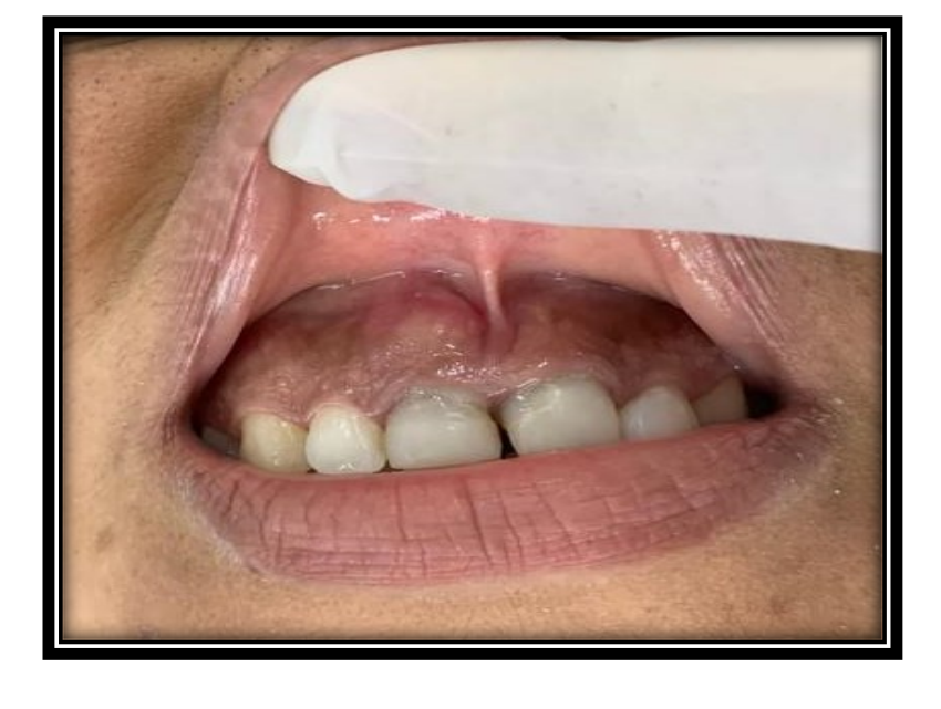
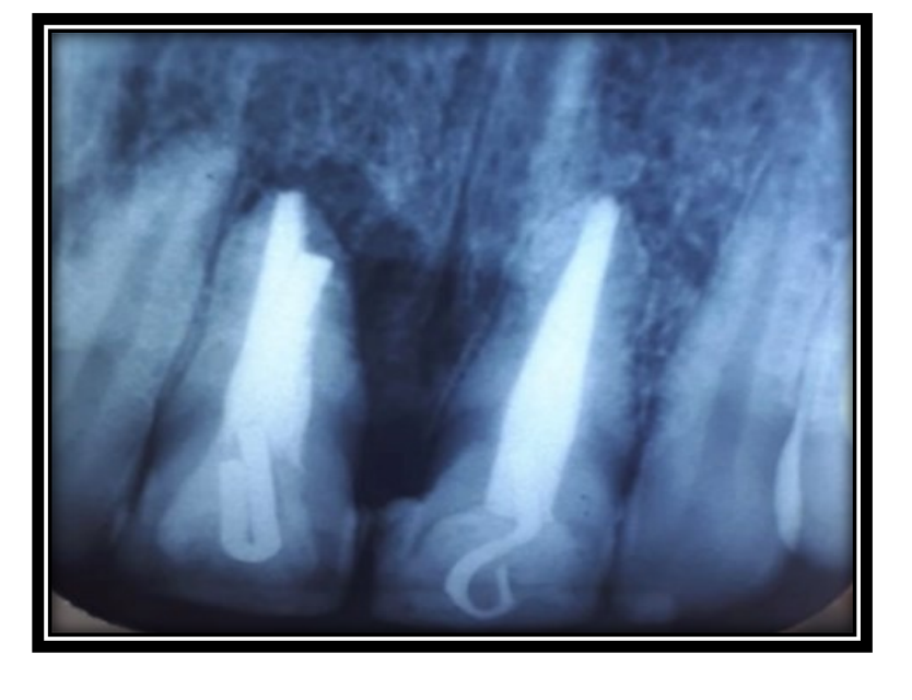
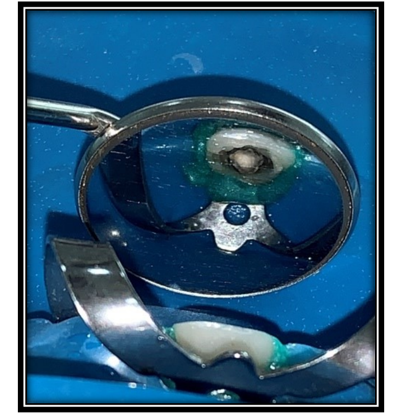
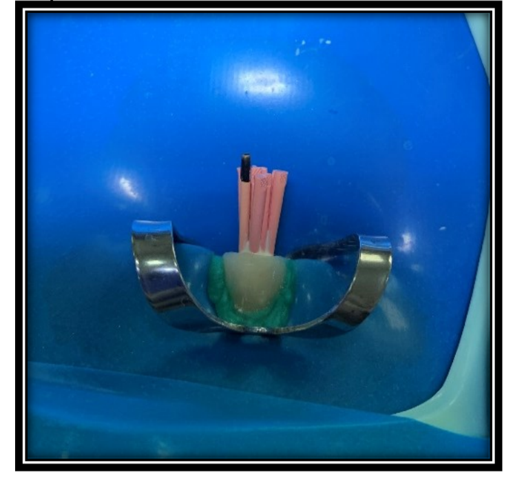
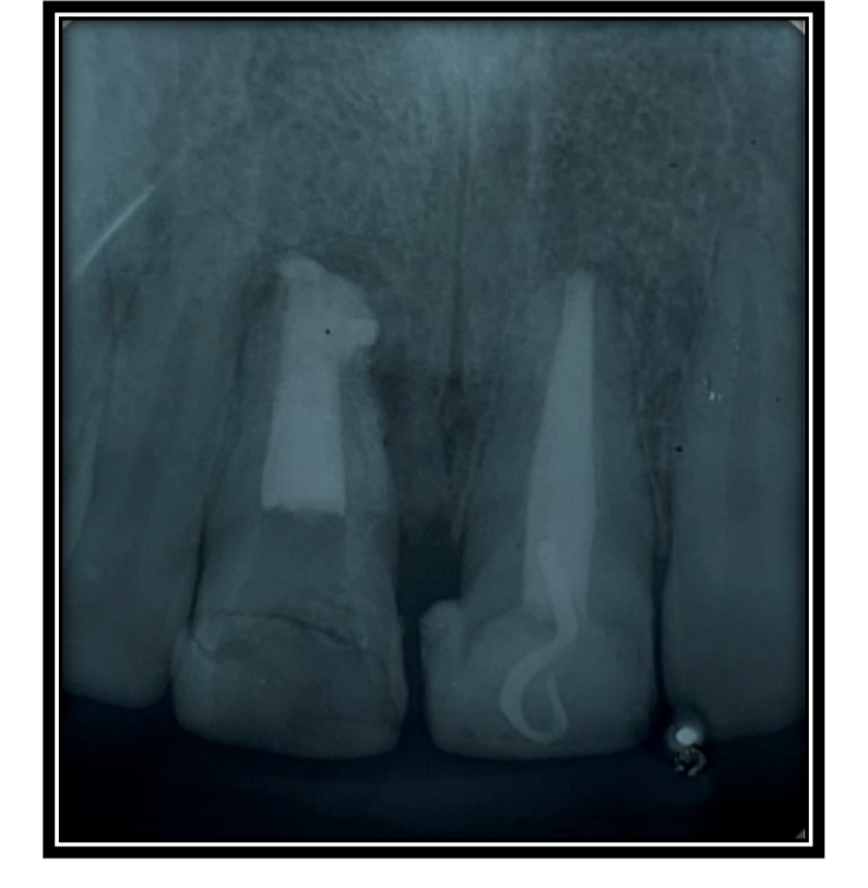
Il paziente MFBN, una donna di 42 anni con melanoderma, ha cercato trattamento presso la clinica dentale di una facoltà di Odontoiatria situata nel Ceará, riferendo una storia di trauma al dente 11 con precedente intervento endodontico. L'esame clinico intraorale ha rivelato un oscuramento del dente, accompagnato da edema ed essudazione (Figura 1). La successiva valutazione radiografica periapicale ha rivelato radiolucenza periapicale suggestiva di perforazione radicolare nella regione apicale (Figura 2). Per confermare la diagnosi di perforazione radicolare apicale, è stato eseguito un esame tomografico.
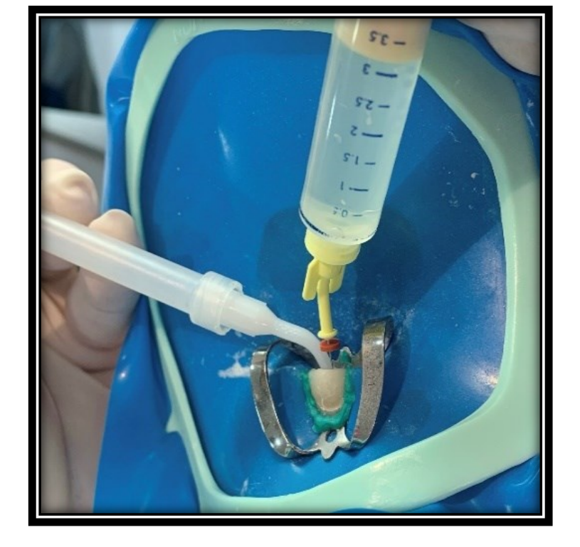
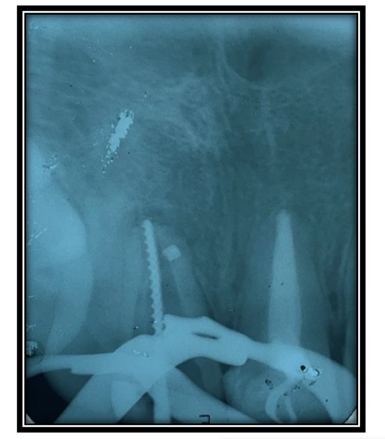
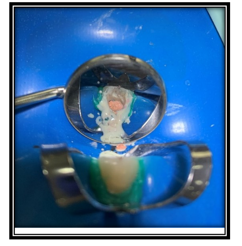
Durante la prima sessione di trattamento, è stata somministrata un'anestesia infiltrativa del nervo alveolare anteriore utilizzando lidocaina al 2% con epinefrina 1:100.000 (DLF, Rio de Janeiro, Brasile) nel dente 11. Sotto isolamento con diga di gomma, la clip utilizzata come ritenzione per la protesi fissa è stata rimossa utilizzando una punta diamantata 1014 (KG-Sorensen, São Paulo, Brasile). Il canale radicolare è stato successivamente pulito utilizzando un file Reciproc R25 e un inserto ultrasonico Clear Sonic (HELSE), supportato da un microscopio elettronico e abbondante irrigazione con ipoclorito di sodio al 2,5% (Figura 3).
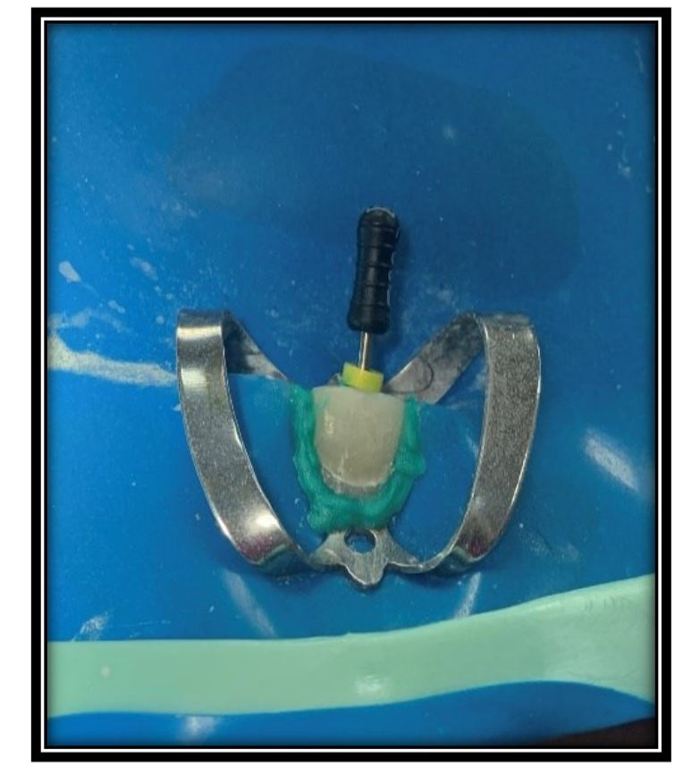
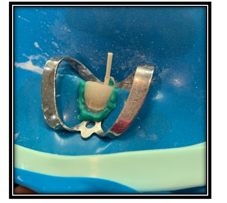
Un file 80 è stato utilizzato per determinare radiograficamente la lunghezza di lavoro (CRT) e eseguire l'istruzione del canale radicolare (Figure 4 e 5). Dopo la preparazione biomeccanica, il cemento riparativo Bio-C Repair è stato inserito con cura nel sito della perforazione utilizzando un punto di carta assorbente (Figura 6). Successivamente, il canale è stato riempito con Calen plus PMCC e è stata applicata una restaurazione temporanea. È stata eseguita una radiografia periapicale di follow-up per confermare il posizionamento preciso del cemento riparativo all'interno della perforazione (Figura 7).
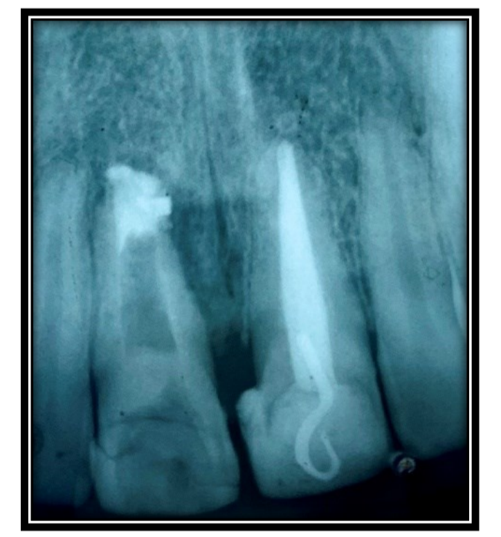
Nella seconda seduta di trattamento, la medicazione intracanalare è stata rimossa utilizzando ipoclorito di sodio al 2,5% attraverso l'Irrigazione Ultrasonica Passiva (PUI). L'irrigazione successiva è stata effettuata con EDTA e ipoclorito di sodio, attenendosi al protocollo finale di irrigazione. Il canale è stato accuratamente asciugato utilizzando punte capillari e coni di carta assorbente (Figura 8). È stato impiegato il sigillante AH Plus per il riempimento del canale, e la tecnica di compattazione verticale a caldo è stata applicata con l'ausilio di McSpadden a bassa rotazione (Figure 9 e 10). Infine, è stata effettuata una radiografia per verificare la qualità del riempimento del canale radicolare (Figura 11).
Discussione
La perforazione radicolare rappresenta una causa prevalente di incidenti operatori, come evidenziato da Alves et al.. È caratterizzata da una comunicazione anomala tra i canali radicolari e i tessuti periradicolari, derivante da fattori patologici o iatrogeni.
Estrela et al. sottolineano l'importanza dei risultati clinici e degli esami radiografici per una diagnosi precisa e la pianificazione del caso. Sebbene la radiografia periapicale sia la tecnica di imaging comunemente utilizzata per diagnosticare la perforazione radicolare, potrebbe mancare di specificità nei casi complessi. In questo contesto, la tomografia offre una moltitudine di vantaggi rispetto alla radiografia tradizionale.
I localizzatori apicali elettronici si sono dimostrati preziosi nella diagnosi delle perforazioni radicolari. Uno studio in vitro di De Miranda Candeiro et al. ha valutato tre localizzatori apicali per la loro accuratezza nel localizzare perforazioni radicolari simulate, dimostrando la loro efficienza nell'aiutare la diagnosi di tali complicazioni.
Il trattamento delle perforazioni radicolari comporta l'identificazione dell'area interessata, seguita dalla decontaminazione e dalla sigillatura appropriata con materiali idonei. La prognosi tende a essere meno favorevole quando le perforazioni si trovano nei terzi medio e apicale, rispetto al terzo cervicale e al pavimento della camera pulpare. Un trattamento precoce gioca un ruolo cruciale nell'influenzare positivamente la prognosi, rendendo la terapia convenzionale l'opzione preferita per risolvere tali casi.
I materiali di sigillatura impiegati per le perforazioni radicolari dovrebbero possedere caratteristiche specifiche, tra cui biocompatibilità, non citotossicità, radiopacità, proprietà battericide, idrofili, stabilità dimensionale, buona capacità di sigillatura e la capacità di prevenire microperdite, come sottolineato da. L'Aggregato Minerale di Triossido (MTA) è stato ampiamente utilizzato nel trattamento delle perforazioni grazie alla sua biocompatibilità, eccellente capacità di sigillatura e proprietà rigenerative dei tessuti, derivanti dalle sue proprietà antimicrobiche e dal pH alcalino (12,5). Tuttavia, i nuovi cementi bioceramici riparativi offrono numerosi vantaggi per risolvere casi complessi di perforazione radicolare.
Attualmente, i cementi bioceramici riparativi sono considerati lo standard d'oro per i casi di perforazione radicolare, mostrando caratteristiche come una capacità di sigillatura efficace, eccellente tolleranza da parte dei tessuti parodontali e l'induzione della riparazione ossea. La loro biocompatibilità è conferita dalla loro somiglianza all'idrossiapatite biologica, come notato da Estrela et al. [8] e Brait et al. Nonostante le loro eccezionali proprietà e vantaggi, l'utilizzo limitato è attribuito all'alto costo dei materiali disponibili in commercio.
Questo studio ha impiegato Bio-C Repair (Angelus), un bioceramico riparativo, che ha dimostrato risultati favorevoli nel trattamento delle perforazioni radicolari intraosseose, mostrando la sua eccellente biocompatibilità, capacità di indurre la riparazione ossea e comodità clinica.
Conclusioni
In conclusione, l'applicazione di cementi bioceramici riparativi, in congiunzione con un robusto protocollo chimico-meccanico, si dimostra efficace nel trattamento delle perforazioni radicolari. L'uso di Bio-C Repair favorisce il recupero dei tessuti periradicolari, facilitando così il ripristino funzionale del dente colpito.
Andressa C. F. de Brito, Clara I. A. S. e Silva, Alyssa M. Pinheiro, Irvina C. F. Melo, Ana B. H. R. D. de Sampaio, Eliane M. G. M. de Vasconcelo, Mario F. de P. Leonardi, Cicero L. G. Ramalho
Riferimenti
- Estrela, C.; et al. Perforazioni radicolari: una revisione di diagnosi, prognosi e materiali. Brazilian oral research, 2018, v. 32, p. 1-15.
- Alzahrani, O.; Alghamdi, F. Gestione non chirurgica della perforazione radicolare apicale utilizzando l'aggregato di triossido minerale. Case reports in dentistry, 2021, v. 2021, p. 1-5.
- Alves, R.A.A.; et al. Un approccio conservativo alla gestione chirurgica della perforazione del canale radicolare. Case reports in dentistry, 2021, v. 2021, p. 1-6. 51, n. 3 Suppl 1, p. S128.
- Sousa, A.S.; Lima, H.M.; Salomão, M.B. CIMENTI MTA E BIOCERAMICI: REVISIONE DELLA LETTERATURA. Revista Cathedral, 2020, v. 2, n. 3, p. 64-74.
- Costa, G.C. Aggregato di triossido minerale: proprietà fisiche, chimiche, biologiche e il suo uso come materiale sigillante in casi di perforazione radicolare: revisione della letteratura. 2021.
- de Miranda Candeiro, G.T.; et al. Trattamento di perforazione radicolare cervicale con l'uso di MTA - rapporto di caso. Research, Society and Development, 2022, v. 11, n. 2, p. e8911225474-e8911225474.
- de França, G.M.; et al. Uso dei bioceramici nell'endodonzia: revisione della letteratura. Revista de Ciências da Saúde Nova Esperança, 2019, v. 17, n. 2, p. 45-55.
- Braitt, A.H.; et al. Sigillatura di perforazione con tecnica di inserimento modificata del MTA: rapporto di caso clinico. Rev. Odontol. Araçatuba (Online), 2022, p. 18-23.
- Arantes, E.A.; Boer, N.C.P. MATERIALI UTILIZZATI NEI TRATTAMENTI DELLE PERFURAZIONI RADICOLARI: REVISIONE DELLA LETTERATURA. Revista Ibero-Americana de Humanidades, Ciências e Educação, 2022, v. 8, n. 5, p. 567-576.
- de Oliveira Resende, F.; et al. Trattamento con MTA Repair HP di una vasta perforazione radicolare dopo iatrogenia: rapporto di caso. Revista da Faculdade de Odontologia-UPF, 2019, v. 24, n. 1, p. 120-126.
- Fehlberg, B.K.; Bittencourt, G. Chirurgia parendodontica—apicoectomia e otturazione simultanea dei canali radicolari con aggregato di triossido minerale (MTA): rapporto di caso. Dental Press Endodontics, 2019, v. 9, n. 1, p. 48-57.
- Raghavendra, S.S.; et al. Bioceramici in endodonzia–una revisione. Journal of Istanbul University Faculty of Dentistry, 2017, v.